Research Article
Association between cadmium concentrations in blood, thyroid tissue and stage of thyroid cancer
Asociación entre las concentraciones de cadmio en sangre, tejido tiroideo y el estadio del cáncer de tiroides
Lam Nguyen Nha Truc1 https://orcid.org/0009-0005-0578-4945
Nguyen Thi Thu Trang2 https://orcid.org/0000-0002-4859-8991
Le Tuan Anh2 https://orcid.org/0000-0001-6288-9077
Hoang Thi Truong2 https://orcid.org/0000-0001-7118-7122
Nguyen Van Chuyen2* https://orcid.org/0000-0001-9507-8377
Nguyen Van Bang3 https://orcid.org/0000-0002-3542-9929
Nguyen Thi Minh Ngoc4 https://orcid.org/0000-0002-7652-659X
Tran Quang Phuc5 https://orcid.org/0009-0009-5661-9770
Nguyen Van Ba6 https://orcid.org/0000-0001-8603-2035
Ta Quang Thanh7 https://orcid.org/0009-0004-0544-808X
1Cho Ray Hospital. Health Care Department. Ho Chi Minh City, Vietnam.
2Vietnam Military Medical University. Department of Military Hygiene. Hanoi, Vietnam.
3Military hospital 103. Department of Hematology. Toxicology Radiation and Occupational Diseases. Hanoi, Vietnam.
4 Haiphong University of Medicine and Pharmacy. Faculty of Public Health. Haiphong, Vietnam.
5The National Institute of Malariology. Parasitology and Entomology. Department of Planning. Hanoi, Vietnam.
6Military hospital 175. Oncology – Nuclear Medicine Institute. Ho Chi Minh City, Vietnam.
7Bac Thang Long hospital. Department of Cardiology – Geriatrics. Hanoi, Vietnam.
*Author for correspondence. Email: nguyenvanchuyenk40@gmail.com
ABSTRACTS
Introduction: The etiology of thyroid cancer remains unclear. Previous studies have supported the hypothesis that cadmium, may contribute to the development of thyroid cancer.
Objectives: To assess cadmium concentrations in thyroid tissue and blood samples, and to evaluate the association between these concentrations and thyroid cancer state.
Methods: A descriptive cross-sectional study was conducted on 282 patients who underwent partial or total thyroidectomy at Cho Ray Hospital. Cadmium concentrations in blood and tissue were determined. The association between Cadmium in blood, thyroid tissue and thyroid cancer stage was assessed by a logistic regression model.
Results: Cadmium levels in blood and tissue samples were higher in patients with cancer tumor node metastasis stage (TNM) stage 2 and 4 compared to stage 1. Logistic regression analysis showed that blood cadmium in the TNM stage 2 and 4 groups were significantly higher than those in stage 1 group (OR = 1.355; 95% CI: 1.176–1.56; p < 0.001). Elevated cadmium levels in thyroid tissue were also positively associated with TNM stages 2 and 4 compared to stage 1 (OR = 1.005; 95% CI: 1.001–1.008; p = 0.007).
Conclusion: Cadmium concentrations in thyroid tissue and blood samples are associated with thyroid cancer stage.
Keywords: association; cadmium; thyroid cancer.
RESUMEN
Introducción: La etiología del cáncer de tiroides sigue siendo incierta. Estudios previos han respaldado la hipótesis de que el cadmio podría contribuir a su desarrollo.
Objetivos: Evaluar las concentraciones de cadmio en el tejido tiroideo y en muestras de sangre, y evaluar la asociación entre estas concentraciones y el estado de cáncer de tiroides.
Métodos: Se realizó un estudio descriptivo transversal en 282 pacientes sometidos a tiroidectomía parcial o total en el Hospital Cho Ray. Se determinaron las concentraciones de cadmio en sangre y tejido. La asociación entre el cadmio en sangre, tejido tiroideo y el estadio del cáncer de tiroides se evaluó mediante un modelo de regresión logística.
Resultados: Los niveles de cadmio en muestras de sangre y tejido fueron más altos en pacientes con metástasis ganglionar tumoral (TNM) en estadios 2 y 4 en comparación con el estadio 1. El análisis de regresión logística mostró que el cadmio en sangre en los grupos TNM en estadios 2 y 4 fue significativamente más alto que en el grupo de estadio 1 (OR = 1,355; IC del 95 %: 1,176-1,56; p < 0,001). Los niveles elevados de cadmio en el tejido tiroideo también se asociaron positivamente con los estadios TNM 2 y 4 en comparación con el estadio 1 (OR = 1,005; IC del 95 %: 1,001-1,008; p = 0,007).
Conclusión: Las concentraciones de cadmio en tejido tiroideo y muestras de sangre se asocian con el estadio del cáncer de tiroides.
Palabras clave: asociación; cadmio; cáncer de tiroides.
Received: 12/08/2025
Approved: 06/10/2025
INTRODUCTION
Thyroid cancer is one of the most common endocrine malignancies worldwide. In Vietnam, the number of thyroid cancer cases in Hanoi and Ho Chi Minh City is projected to increase from 2,020 cases in 2020 to 2,971 cases by 2025, making it one of the five most prevalent cancers in the country by 2025(1). Thyroid cancer is associated with a significant economic burden and reduced quality of life, making it an important national health issue. Various efforts have been made to identify the causes of thyroid cancer. Although radiation exposure, elevated thyroid-stimulating hormone levels, overweight, insulin resistance, and environmental pollution have been identified as risk factors for thyroid cancer, these conclusions have yet to be definitively confirmed. Identifying potential contributing factors to the development of thyroid cancer is therefore of great importance.
Cadmium (Cd) is a common toxic heavy metal, known as an endocrine disruptor affecting the thyroid gland and a carcinogen. The International Agency for Research on Cancer (IARC) classifies cadmium as a human carcinogen (2). A study conducted in South Korea demonstrated an association between heavy metal concentrations in blood and tissue and the staging of thyroid cancer in women(3). Therefore, cadmium (Cd) has become a major focus in research on the causes of thyroid cancer. In Vietnam, the association between thyroid cancer staging and Cd concentrations in tissue and blood has not been extensively analyzed. Thus, this study aims to compare Cd levels between patients with malignant thyroid nodules and those with benign thyroid nodules. Additionally, the relationship between Cd concentrations in thyroid tissue and blood samples of thyroid cancer patients and the stage of thyroid cancer were analyzed.
METHODS
Study Design
This study was a descriptive cross-sectional study with control. The Cd concentrations in blood, thyroid tissue of the case and control groups at the time of the study were compared. In addition, the relationship between Cd in blood, thyroid tissue and thyroid cancer stage was analyzed.
Study Participants
Study subjects were selected from October 1, 2024, to April 30, 2025, at Cho Ray Hospital.
Inclusion criteria
For the case group, inclusion criteria for case groups were patients presenting with clinical suspicion of thyroid nodules, who underwent thyroid ultrasound and were classified as Thyroid Imaging, Reporting and Data System (TIRADS) 4 or 5. Patients who underwent partial or total thyroidectomy with nodules, and postoperative histopathological results confirmed carcinoma were selected for the disease group (n = 217). At the time of diagnosis, the patient had not received any specific treatment. The patients agreed to participate in the study.
For the control group, inclusion criteria were patients presenting with clinical suspicion of thyroid nodules, who underwent thyroid ultrasound and were classified as Thyroid Imaging, Reporting and Data System (TI-RADS) 4 or 5.(4) Patients who underwent partial or total thyroidectomy with nodules, with postoperative histopathology confirming benign lesions were selected for the control group (n = 65). The patients agreed to participate in the study.
Exclusion criteria
Cases of secondary tumors (tumors not originating from the thyroid gland but metastasizing from other regional tumors); patients with other thyroid diseases such as acute thyroiditis, active Basedow’s disease, hyperthyroidism or hypothyroidism; individuals with a history of any chronic diseases including chronic liver disease, liver dysfunction, cardiovascular diseases, rheumatic, neurological, and/or autoimmune disorders; pregnant or breastfeeding women were excluded. Ultimately, 217 patients with postoperative histopathological diagnosis of Carcinoma were assigned to the case group, while 65 patients with benign histopathological results were selected as the control group.
Variables
Analytic variables included: Blood Cd; Tissue Cd; TNM staging (AJCC 8th edition); T (tumor); N (lymph node); M (metastasis).
Analysis of heavy metals
Chemicals
Standard solutions of Cd, (1000 ppm) were purchased from Merck (Darmstadt, Germany). Sulfuric acid (H2SO4 98%), nitric acid (HNO3 65%), hydrogen peroxide (H2O2), perchloric acid (HClO4 70%) were also purchased from Merck. All of the chemical solutions were freshly prepared each day. The standard curve solutions were diluted with HNO3 (1%).
Thyroid tissue and blood collection
To analyze heavy metals in blood and thyroid tissue samples, specimens were collected as follows: 2 mL of venous blood was drawn into tubes containing heparin as an anticoagulant. For tissue samples, approximately 0.5 grams of normal thyroid tissue surrounding the nodule was obtained during surgery, wrapped in sterile gauze, and placed into 50 mL Falcon tubes containing physiological saline. After collection, samples were stored at temperatures between 0 and 4 °C in sample storage boxes, then immediately transported to the laboratory and stored at 20 °C until analysis. Blood cadmium concentrations were compared with the reference values from ISTISAN 10/22.(5)
Microwave sample preparation
Accurately weighed 0.5 g of thyroid tissue sample and 2 mL of blood sample. The thyroid tissue and blood samples were digested using a mixture of HNO₃, H₂SO₄, and HClO₄ in a ratio of 10:1:1, combined with 5 mL of H₂O₂, in the Multiwave PRO microwave reaction system (Anton Paar GmbH, Graz, Austria).
First, samples were placed into Teflon digestion tubes, and then the digestion mixture (HNO₃, H₂SO₄, HClO₄, H₂O₂) was added. After gentle shaking for 20 minutes to initiate the digestion reaction, the tubes were placed in the Multiwave PRO system to complete sample digestion (USEPA, 1996). The temperature program was set as follows: Heating to 170 °C for 5 minutes and holding for 10 minutes; then increasing to 200 °C and holding for 15 minutes, followed by cooling to room temperature. After digestion, the sample solution was expected to be clear and transparent. The digested samples were then diluted with 1% HNO₃ to a final volume of 5 mL prior to analysis. The dilution factor for cadmium (Cd) was calculated accordingly 5.
Analysis of Cd
Heavy metals in the samples were analyzed at the Laboratory of the Department of Military Hygiene, Vietnam Military Medical University.
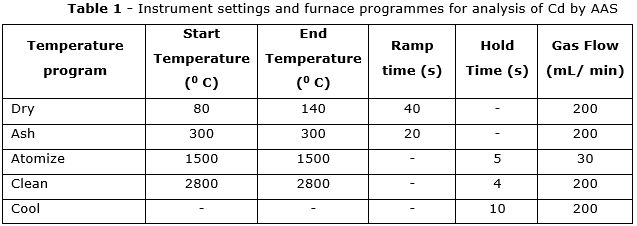
Atomic Absorption Spectrophotometry (AAS) (Model AZ3000, Hitachi Ltd., Tokyo, Japan) was used for quantification. Cadmium (Cd) was measured using the graphite furnace method at a wavelength of 228.8 nm, slit width of 1.3 nm, and lamp current of 7.5 mA. The limit of detection (LOD) for Cd was 1 ppb. A standard calibration curve was constructed using at least five concentration points for each element. The conditions and temperature programs used for determining Cd levels are detailed in table 1.
The standard concentrations for cadmium (Cd) were 0, 1, 5, 10, and 20 ppb. A correlation coefficient of 99.99% indicated a highly linear relationship across the studied concentration range.
To check the accuracy of the method, the percentage recoveries were analyzed and quantified. The recovery coefficients for in samples of thyroid tissue and blood after adding the spiked sample were calculated, ranging from 94.05% to 95.06% for Cd. The values of these recovery coefficients indicated that the method was highly accurate and could be used for the analysis of heavy metals.
Histopathological examination and TNM staging of thyroid cancer
Details of thyroid surgery included partial or total thyroidectomy, histopathological results (tumor size), and TNM staging. Tumor size was defined as the largest diameter of the carcinoma based on postoperative biopsy results. All patients were staged according to the TNM classification (tumor/lymph node/metastasis) following the American Joint Committee on Cancer (AJCC) 8th edition.(6)
Statistical analysis
The concentrations of heavy metals were expressed based on five replicate measurements of Cd. Data were analyzed using one-way analysis of variance (ANOVA). All statistical analyses were performed with SPSS software (version 19.0, IBM Corp., Armonk, NY, USA). The results of Cd in blood and thyroid tissue samples were presented as mean [standard deviation (SD)] and minimum and maximum values. The categorical variables are expressed as numbers and percentages of total subjects. Logistic regression analysis was conducted to estimate odds ratios (OR) and 95% confidence intervals (CI) for the association between Cd concentrations in tissue and blood samples of thyroid cancer patients and cancer staging. A p-value < 0.05 was considered statistically significant.
Ethical considerations
This study was approved by the Ethics Committee under Decision No. 1178/QĐ-VSR of the National Institute of Malariology, Parasitology and Entomology on December 8, 2023, and by the Ethics Committee for Biomedical Research under approval No. 1912/GCN-HĐĐĐ dated December 3, 2024, of Cho Ray Hospital.
RESULTS
Histopathological results and staging of thyroid cancer
Table 2 shows that among the 282 study subjects, 217 patients were diagnosed with thyroid cancer. When categorized by the TNM staging system for thyroid cancer, the most common stage was stage 1, accounting for 170 patients (78.34%), followed by stage 2 and stage 4, representing 21.19% and 0.47%, respectively. No patients were classified in stage 3.
Furthermore, the T (tumor) staging results indicated that most patients were at stage T3, comprising 116 cases (53.45%), while 89 patients (41.01%) were at stage T1. The N (lymph node) staging showed that 34 patients (15.66%) had lymph node metastasis (N1). There was one patient (0.5%) with distant metastasis (M1).
The comparison of Cd concentrations in thyroid tissue and blood samples of thyroid cancer patients and control group
Table 3 compares the mean cadmium (Cd) concentrations in tissue and blood samples between the case group and the control group. There was a significant difference in Cd levels between the two groups. The mean blood Cd concentration in thyroid cancer patients was 1.75 μg/L (n = 217), whereas in the control group it was 1.27 μg/L (n = 65). While 31.33% of thyroid cancer patients had blood Cd concentrations exceeding the permissible limit, only 4.60% of the control group exceeded this threshold. Additionally, the mean Cd concentration in tissue samples was 165.95 μg/kg (weight) for the thyroid cancer group and 161.79 μg/kg (weight) for the control group.
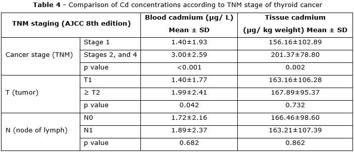
Comparison of Cd concentrations in thyroid tissue and blood samples by thyroid cancer stage
The comparison of Cd concentrations in blood and tissue samples across thyroid cancer stages is presented in table 4. Blood Cd levels in patients at TNM stage 1 were significantly lower than those in stages 2 and 4, with mean values of 1.40 μg/L and 3.00 μg/L, respectively (p < 0.001). Comparing Cd levels by T stage showed that the ≥ T2 group had significantly higher blood Cd concentrations than the T1 group, with means of 1.99 μg/L and 1.40 μg/L, respectively (p < 0.05). There was no statistically significant difference in blood Cd concentration between N0 and N1 groups. Cd concentrations in tissue samples were higher in TNM stages 2 and 4 compared to stage 1 (p < 0.002). However, no significant difference was observed in tissue Cd levels between T1 and ≥ T2 groups.
Odds ratios (OR) for Cd concentrations in tissue and blood samples by thyroid cancer stage
Based on the statistically significant Cd concentrations shown in table 4, the association between heavy metal levels in tissue and blood samples and thyroid cancer stages were analyzed (table 5). Logistic regression analysis revealed that blood Cd concentrations were higher in the TNM stage 2 and 4 groups compared to the TNM stage 1 group (OR = 1.355; 95% CI: 1.176–1.56; p < 0.001). For the progressive tumor stage group (≥ T2), logistic regression showed higher blood Cd concentrations compared to the T1 group, although this difference was marginally non-significant (OR = 1.137; 95% CI: 0.997–1.297; p = 0.056). High Cd concentrations in tissue samples were positively associated with TNM stages 2 and 4 compared to stage 1 (OR = 1.005; 95% CI: 1.001–1.008; p = 0.007)
DISCUSSION
Histopathological findings and thyroid cancer staging
According to the TNM classification, this study included 89 patients at stage T1, and only 7 patients at stage T4. The numbers of patients at stages T2 and T3 were 5 and 116, respectively. The number of patients without lymph node metastasis (N0) and with lymph node metastasis (N1) were 183 and 34, respectively. There was one patient diagnosed with distant metastasis (M1).
Park E et al.(3) found that chronic accumulation of Cd in thyroid tissue promotes the progression of thyroid cancer. In this study, the mean Cd concentrations in thyroid tissue for the TNM stage 1 group and the combined TNM stages 2 and 4 group were 156.16 and 201.37 µg/kg, respectively (p = 0.002). Similarly, Cd levels in blood samples were significantly higher in TNM stages 2 and 4 compared to stage 1 (p < 0.001). It is somewhat difficult to compare these results with previous studies conducted in Vietnam due to the limited number of reports investigating the relationship between Cd concentrations in tissue samples and thyroid cancer. Therefore, this study was compared to several international studies.
High concentrations of Cd in blood and tissue may be one of the contributing factors to the development of thyroid cancer. The thyroid gland is an organ where Cd can accumulate throughout a person's lifetime. This accumulation may lead to various thyroid dysfunctions. Therefore, Cd levels in blood and tissue are important factors that warrant thorough investigation.
In this study, Cd concentrations in tissue and blood samples between subjects with histopathological results of malignant thyroid nodules and those with benign thyroid nodules were compared. Cd is more toxic than Pb and Hg, and is classified as a human carcinogen (2). These results indicate that blood Cd concentrations in the thyroid cancer group were higher than those in the control group. These findings are consistent with the conclusions of several previous studies. However, the mean blood Cd concentration in this study was lower than that reported by Shao R et al.(7) (2.085 μg/L), which analyzed heavy metal concentrations in patients with hyperthyroidism, hypothyroidism, and thyroid cancer. They reported that Cd concentrations were higher in the disease group compared to the control group. In current study, Cd concentrations in thyroid tissue were also higher in the cancer group than in the control group. These findings suggest that Cd may be a potential carcinogenic factor for the thyroid gland.
Comparison of heavy metal concentrations by thyroid cancer stage
Different stages of thyroid cancer and the concentrations of heavy metals in blood and tissue samples were analyzed in this study. Cd concentrations in both blood and tissue showed a positive correlation with thyroid cancer stages as classified by the TNM staging system. Compared to patients with TNM stage 1 disease, those at stages 2 and 4 had higher Cd levels in both tissue and blood samples. Therefore, in this study, Cd may be considered a potential risk factor contributing to the progression of thyroid cancer. Comparison between this findings and previous international studies also suggests a correlation between Cd concentrations in the blood and tissue of thyroid cancer patients and the stage of the disease.(8,9)
A similar conclusion was also demonstrated by He JL et al.(10) in their study on patients with thyroid cancer. Their findings indicate that more advanced cancer stages are associated with higher Cd concentrations, and confirmed a positive association between thyroid cancer and urinary Cd levels.
Based on the above findings, it can be shown that there is a relationship between cadmium (Cd) accumulation and the progression of thyroid cancer. The toxic effects of Cd on thyroid function may occur at both the cellular and peripheral levels through various mechanisms. At the cellular level, excessive accumulation of cadmium in the thyroid gland may affect transcriptional activity or cellular signaling pathways, leading to activation of mitogenic cascades such as proto-oncogene expression, DNA synthesis, and cell proliferation, thereby acting as a potential carcinogen. At the peripheral level, Cd has been shown to disrupt thyroid hormone homeostasis (11). In this study, Cd concentrations in both tissue and blood were positively associated with thyroid cancer stage. This may reflect that Cd could be a contributing factor in the progression or aggravation of thyroid cancer.
The main of this study is its cross-sectional design. To our knowledge, this is among the first studies in Vietnam to concurrently measure cadmium in blood and thyroid tissue in relation to thyroid cancer stage. In summary, Cd concentration in the cancer group was higher than that in the control group. This study found that high cadmium (Cd) concentrations in both blood and thyroid tissue were associated with more advanced stages of thyroid cancer, as classified by the TNM staging system.
Cadmium concentrations in thyroid tissue and blood samples are associated with thyroid cancer stage.
BIBLIOGRAPHIC REFERENCES
1. Nguyen S.M, Deppen S, Nguyen G.H, Pham D.X, Bui T.D. Projecting cancer incidence for 2025 in the 2 largest populated cities in Vietnam [Internet]. Cancer control. 2019; 26(1): 1073274819865274. DOI: https://doi.org/10.1177/1073274819865274
2. IARC Working Group on the Evaluation of Carcinogenic Risks to Humans. Beryllium, cadmium, mercury, and exposures in the glass manufacturing industry [Internet]. Lyon (FR): International Agency for Research on Cancer; 1993. [access: 12/08/2025]. Available from: https://www.ncbi.nlm.nih.gov/books/NBK499756/
3. Park E, Kim S, Song SH, Lee CW, Kwon JT, Lim MK, et al. Environmental exposure to cadmium and risk of thyroid cancer from national industrial complex areas: a population-based cohort study [Internet]. Chemosphere. 2021; 268: 128819. DOI: https://doi.org/10.1016/j.chemosphere.2020.128819
4. Tessler FN, Middleton WD, Grant EG, Hoang JK, Berland LL, Teefey SA, et al. ACR Thyroid Imaging, Reporting and Data System (TI-RADS): White Paper of the ACR TI-RADS Committee. J Am Coll Radiol. 2017;14(5):587–595. DOI: https://doi.org/10.1016/j.jacr.2017.01.046
5. Ruffolo P, Acquaviva O, Capece P, Mazzei F, Ruffolo B, Panunzio M, et al. From the Histological Model to the Mutational Model: The Study of Heavy Metals and Other Substances in New Antineoplastic Therapies [Internet]. Glob J Med Res. 2023; 23: 7-9. DOI: https://doi.org/10.34257/GJMRBVOL23IS1PG7
6. Erazo-Puentes MC, Sánchez-Torres A, Aguirre-Urizar JM, Bara-Casaus J, Gay-Escoda C. Has the 8th American joint committee on cancer TNM staging improved prognostic performance in oral cancer? A systematic review [Internet]. Medicina Oral, Patología Oral y Cirugía Bucal. 2024; 29(2): e163. DOI: https://doi.org/10.4317/medoral.25983
7. Shao R, Su L, Wang P, Han X, Wang T, Dai J, et al. Higher cadmium exposure was associated with sex-specific thyroid dysfunction: Consistent evidence from two independent cross-sectional studies based on urinary and blood cadmium measurements [Internet]. Research Square [preprint]. 2023: rs. 3. rs-3455102. DOI: https://doi.org/10.21203/rs.3.rs-3455102/v1
8. Chung HK, Nam JS, Ahn CW, Lee YS, Kim KR. Some elements in thyroid tissue are associated with more advanced stage of thyroid cancer in Korean women, Biol. Trace Elem. Res. 2016; 171: 54–62. DOI: https://doi.org/10.1007/s12011-015-0502-5
9. Zhang C, Hua-Bing W, Meng-Xia C. Association of exposure to multiple metals with papillary thyroid cancer risk in China. Environmental Science and Pollution Research. 2019; 26:20560–572. DOI: https://doi.org/10.1007/s11356-019-04733-x
10. He JL, Wu HB, Hu WL, Liu JJ, Zhang Q, Xiao W, et al. Exposure to multiple trace elements and thyroid cancer risk in Chinese adults: A case-control study [Internet]. International Journal of Hygiene and Environmental Health. 2022; 246: 114049. DOI: https://doi.org/10.1016/j.ijheh.2022.114049
11. Capriglione F, Maiuolo J, Celano M, Damante G, Russo D, Bulotta S, et al. Quercetin protects human thyroid cells against cadmium toxicity [Internet]. International Journal of Molecular Sciences. 2021; 22(13): 6849. DOI: https://doi.org/10.3390/ijms22136849
Conflicts of interest
The authors declare that they have no potential conflicts of interest relevant to this article.
Authorship contribution
Conceptualization: Lam Nguyen Nha Truc, Nguyen Van Ba, Tran Quang Phuc.
Data curation: Lam Nguyen Nha Truc, Le Tuan Anh, Nguyen Thi Thu Trang, Hoang Thi Truong. Formal analysis: Lam Nguyen Nha Truc, Nguyen Van Chuyen.Research: Lam Nguyen Nha Truc, Nguyen Thi Minh Ngoc, Nguyen Van Bang, Ta Quang Thanh.
Methodology: Lam Nguyen Nha Truc, Nguyen Thi Minh Ngoc, Nguyen Van Bang, Ta Quang Thanh.
Supervision: Nguyen Van Chuyen.
Drafting - Revision and editing: Lam Nguyen Nha Truc, Nguyen Van Ba, Nguyen Van Chuyen.
Data Availability Statement
The database is available upon request to the corresponding author at the following email address: nguyenvanchuyenk40@gmail.com