Research Article
Variants of the deep femoral artery in adult Vietnamese people: Autopsy based study
Variantes de la arteria femoral profunda en adultos vietnamitas: Estudio basado en autopsias
Doan Duong Chi Thien1 https://orcid.org/0009-0008-9061-7886
Nguyen Hoang Vu2 https://orcid.org/0009-0003-5957-5677
Nguyen Thanh Van1* https://orcid.org/0000-0002-4310-9191
1Tra Vinh University. School of Medicine and Pharmacy. Travinh, Vietnam.
2University of Medicine and Pharmacy. Department of Anatomy. Ho Chi Minh City, Vietnam.
*Author for correspondence. Email: drthanhvan@gmail.com
ABSTRACT
Introduction: Understanding anatomic variations of the deep femoral artery (DFA) and its branches is crucial for diagnostic angiography and surgical procedures.
Objective: To determine the morphological characteristics and anatomical variations of the deep femoral artery in adult Vietnamese individuals.
Methods: Anatomical variations of the DFA and its branches were examined in 79 thighs from 40 adult cadavers at the University of Medicine and Pharmacy, Ho Chi Minh City (2023). Dissection was performed to determine the origin, branching patterns, and distance of DFA from the inguinal ligament (IL).
Results: The DFA was absent in 2.5% of cases. Its origin from the femoral artery was most commonly posterolateral (29.8%) and posterior (40.3%). The mean distance of DFA origin from the IL was 36.92 ± 16.54 mm. High and middle origins occurred in 35.1% and 58.4% of cases, respectively, while low origin was rare (6.5%). High origin was more frequent in females (48.3% vs. 27.1%, p < 0.05). The LCFA arose from the DFA in 83.2% of cases and from the femoral artery in 13%. The MCFA originated from the DFA in 55.6% and from the femoral artery in 36.4%.
Conclusions: The branching variability of the DFA and its circumflex branches is clinically significant for vascular access, orthopedic procedures, and reconstructive surgery. Recognizing these variations may help prevent iatrogenic complications during femoral interventions.
Keywords: anatomic variation; femoral artery; Vietnam.
RESUMEN
Introducción: Comprender las variaciones de la arteria femoral profunda (AFP) y sus ramas es crucial para la angiografía diagnóstica y los procedimientos quirúrgicos.
Objetivo: Determinar las características morfológicas y las variaciones anatómicas de la AFP en adultos vietnamitas.
Métodos: Se examinaron variaciones anatómicas de la AFP y sus ramas en 79 muslos de 40 cadáveres adultos, en la Universidad de Medicina y Farmacia de Ciudad Ho Chi Minh (2023). Se realizaron disecciones para determinar el origen, los patrones de ramificación y la distancia de la AFP al ligamento inguinal (LI).
Resultados: La AFP estuvo ausente en el 2,5 % de los casos. Su origen desde la arteria femoral fue principalmente posterolateral (29,8 %) y posterior (40,3 %). La distancia media desde el LI fue 36,92 ± 16,54 mm. Los orígenes altos y medios ocurrieron en 35,1 % y 58,4 % de los casos, mientras que el origen bajo fue raro (6,5 %). El origen alto fue más frecuente en mujeres (48,3 % vs. 27,1 %, p< 0,05). La arteria circunfleja femoral lateral (ACFL) surgió de la AFP en 83,2 % de los casos y de la femoral en 13 %. La arteria circunfleja femoral medial (ACFM) se originó de la AFP en 55,6 % y de la femoral en 36,4 %.
Conclusiones: La variabilidad de la AFP y sus ramas circunflejas es clínicamente significativa para el acceso vascular, los procedimientos ortopédicos y la cirugía reconstructiva. Reconocer estas variaciones puede ayudar a prevenir complicaciones iatrogénicas durante intervenciones femorales.
Palabras clave: arteria femoral; variación anatómica; Vietnam.
Received: 01/09/2025
Approved: 05/11/2025
INTRODUCTION
Anatomic variations of the deep femoral artery (DFA) constitute a matter of great interest to anatomists, surgeons, and interventional radiologists due to their significant clinical relevance.(1) The DFA is the biggest branch of the lateral or posterior aspect of the common femoral artery (CFA) in the femoral triangle, located 2 - 6 cm below the inguinal ligament.(2) It is the main vessel for the blood supply of the adductors, flexors, and extensors thigh muscles, as well as of the hip joint and the femur.(3) Moreover, it plays a crucial role in the collateral blood flow between the lower pelvis and the infrapopliteal circulation.(4) The major branches of the DFA are the lateral circumflex femoral artery (LCFA) from its lateral aspect and the medial circumflex femoral artery (MCFA) from its medial wall.(5) The varying vascular anatomy of these vessels is of the utmost importance due to their involvement in vascular, orthopedic, and plastic and reconstructive surgery. (6,7,8)
Knowledge of the exact origin of the LCFA is important for surgeons when applying anesthesia to the femoral nerve, in orthopedic surgeries during femoral and hip procedures, when harvesting an anterolateral thigh (ALT) flap in reconstructive surgery, in aorto-popliteal bypass, in extra/intracranial bypass surgeries, but also coronary artery bypass grafting.(1,9,10,11) Knowledge of the MCFA origin and course variations is pivotal when performing both trochanteric and intertrochanteric osteotomies, in a total arthroplasty to avoid iatrogenic avascular necrosis of the head of the femur, and during flap plastic surgery, as well as in interventional radiology during puncture of the femoral artery.(12,13)
Although numerous studies have investigated the variations of the DFA and its branches across different populations, data on the DFA in Vietnamese adults remains limited. Genetic and biological differences among ethnic groups may result in anatomical variations. Therefore, this study aims to determine the morphological characteristics and anatomical variations of the deep femoral artery in adult Vietnamese based on cadaveric samples.
METHODS
Study design and participants
This descriptive cadaveric study was conducted on 79 thighs from 40 adult cadavers (39 with both thighs and 1 with only one thigh) used for teaching at the Department of Anatomy, University of Medicine and Pharmacy, Ho Chi Minh City, Vietnam, in 2023. The cadavers met the following criteria: The inguinal, thigh region and femoral artery were intact; the thigh region was free from deformities, old surgical scars, or previous dissections; the femoral artery was not damaged or ruptured during dissection; and the cadavers did not have any femoral artery-related pathologies such as aneurysms, arteriovenous fistulas, or significant arterial diseases. Since the DFA was absent in two specimens, anatomical variations were analyzed in 77 thighs (48 male, 29 female; 41 right, 38 left). The age of the standard formalin fixed donors ranged from 25 to 92 years.
Implementation method
The dissection started by incision and reflection of the skin. In the next step, fascia lata was incised and the femoral triangle was exposed according to the prescription of Tillmann and Schünke (1993). FA and its branches were exposed. PFA and its medial and lateral branches (MCFA, LCFA) were dissected and identified. The distance of the midpoint of inguinal ligament (IL) to the origin of DFA was measured with an anthropological calliper. Three groups were established due to the height of origin of DFA from FA: 1. High origin, about 1–2 cm below IL, 2. Middle origin, about 3–5 cm below IL, 3. Low origin, about 6–10 cm below IL. The borders of the three classes were given according to the distribution of the observed different origins of DFA from FA (Fig. 1).
Variables
Demographic characteristics: Exact age, sex, and examined limb side.
Presence of arteries (yes/no): Deep femoral artery (DFA), lateral circumflex femoral artery (LCFA), medial circumflex femoral artery (MCFA).
Branching pattern of DFA: First branch, second branch, isolated origin, and other rare variants (additional branches) from DFA, LCFA, or MCFA.
Variables related to the origin of DFA on the femoral artery (FA) included:
Direction of origin (on the cross-section of FA): Posterior, posterolateral, lateral, anterolateral, medial, posteromedial.
Distance from DFA origin to the midpoint of the inguinal ligament (IL) (mm, continuous variable).
Classification of DFA origin level according to the distance from the IL: High origin: 1–2.9 cm, middle origin: 3–5.9 cm, low origin: ≥ 6 cm.
Comparisons: Differences in the position and distance of DFA origin were analyzed according to sex (male/female) and limb side (right/left).
Statistical analysis
All statistical analyses were performed using SPSS version 26.0 (IBM Corp., Armonk, NY, USA). Continuous variables were expressed as mean ± standard deviation (SD) or median (interquartile range, IQR) depending on distribution. Categorical variables were presented as absolute numbers and percentages. Comparisons between groups (sex and side) were conducted using the Mann–Whitney U test for continuous data. When the expected frequency in any cell was less than 5, Fisher’s exact test was applied instead of the Chi-square test. A p-value ≤ 0.05 was considered statistically significant.
Ethical considerations
This study was approved by the ethics committee of the University of Medicine and Pharmacy, Ho Chi Minh City, Vietnam (Approval No. 3183/ĐHYD-HĐĐĐ) and followed the guidelines of the Declaration of Helsinki.
RESULTS
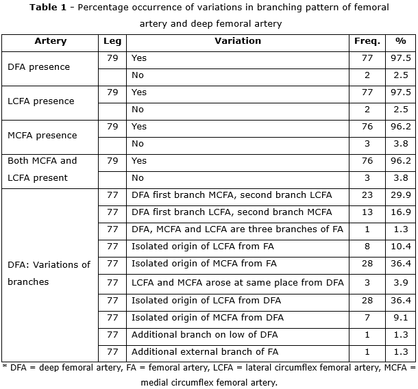
The results showed that the DFA was absent in 2.5% of specimens, while the LCFA and MCFA were absent in 2.5% and 3.8% of cases, respectively (table 1). Among the 77 thighs with a present DFA, the most frequent branching variations included DFA giving rise first to the MCFA followed by the LCFA (29.9%) or, conversely, LCFA first then MCFA (16.9%). Isolated origins of circumflex arteries were also common, with the LCFA arising independently from the FA in 10.4% and from the DFA in 36.4% of cases, while the MCFA originated from the FA in 36.4% and from the DFA in 9.1%. Less frequent variants included trifurcation of the FA into DFA, LCFA, and MCFA (1.3%) and a common trunk for LCFA and MCFA from the DFA (3.9%). Rare additional branches were observed in isolated cases (1.3% each).
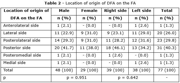
The DFA primarily originated from the posterior to posterolateral region of the FA (table 2): Posterior (31 cases, accounting for 40.3%), posterolateral (23 cases, accounting for 29.8%), and lateral (20 cases, accounting for 26.0%). It was rare for the DFA to originate from the anterior or medial aspects of the FA (1 case from the anterolateral side, 1 case from the medial side, and 1 case from the posteromedial side). The origin sites of DFA on FA showed no significant differences between sexes, sides (p > 0.05).
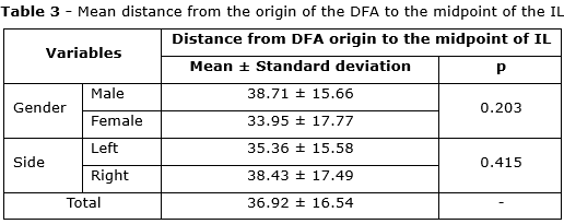
The mean distance from the origin of the DFA to the midpoint of the IL was 36.92 ± 16.54 mm (table 3), with no significant differences observed between males and females, or between the right and left sides (p > 0.05).
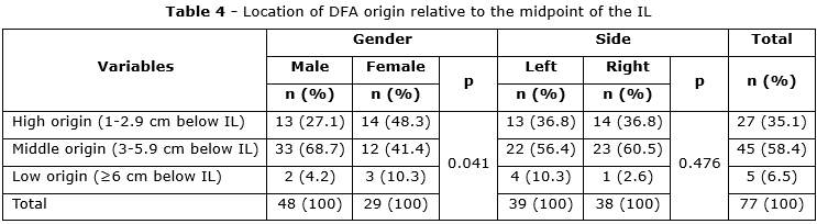
All DFA origins were located below the IL.(table 4) The majority had a middle origin (58.4%), followed by high (35.1%) and low origins (6.5%). High-origin DFA was significantly more frequent in females than in males (p = 0.041), whereas middle-origin DFA was predominant in males. No significant side-to-side difference was observed (p = 0.476).
DISCUSSION
Topography of origin of deep femoral artery from femoral artery
Regarding the origin of the DFA from the FA, the most common sites are the posterolateral and lateral aspects (50-70%), which are more frequent than the posterior and posteromedial origins.(1) In the study by Moaty MA et al.(5) on 20 lower limbs and 92 angiographic cases, the authors reported that the most common origin of the profunda femoris artery (PFA) was from the posterolateral aspect of the femoral artery, accounting for 49.2% on the right side and 62.7% on the left side. A lateral origin was less frequent (33.9% on the right and 23.7% on the left), whereas a posteromedial origin was the least common. This trend is also observed in the present series, where the posterolateral and lateral origins predominate. One point to note is that DFA from the posterior FA (40.3%) is higher than most previous studies,(14) the results of this study are similar to Tzouma G et al.(1) (40.0%) or Manjappa T et al.(15) (40% in the right thigh).
Understanding the DFA's origin site is critical for avoiding iatrogenic femoral arteriovenous fistula during femoral artery punctures.(16) When the DFA is located anterior to the femoral vein, it is very vulnerable during interventional cardiovascular procedures and causes complications of arteriovenous fistula.(17) Variants in the DFA origin, particularly from the anterior aspect of the FA, can lead to complications during angiographic procedures, such as percutaneous transluminal coronary angioplasty, resulting in hematomas or femoral arteriovenous fistulae.(18) Thus, performing an ultrasound prior to angiographic procedures is essential to detect arterial deviations and avoid complications.
Median distance of separation of deep femoral artery from femoral artery in relation to inguinal ligament
The median distance of separation of the DFA from the FA was investigated by several authors.(19,20) Claassen H et al.(14) first proposed a three-level classification of DFA origin depth relative to the inguinal ligament. In the present series of 77 thighs, a median distance of separation of the DFA from the FA in relation to the IL of 3.29 cm was observed. Among these cases, high (1-2 cm below IL) and middle (3-5 cm below IL) origins of the DFA were found in nearly equal distribution (35.1% and 58.4%), while low origins (6-10 cm below the IL) were more seldom (6,5%) (table 2). Interestingly, in the study by Claasen H et al.(14) a trend toward a sex difference was observed (p = 0.096), with deeper origins more frequent in males. This is more evident in the present series: DFA originating at a high position is more common in females than in males (48.3% vs. 27.1%, p < 0.05) and the opposite is true for intermediate origins.
Classifications concerning the variant origins of circumflex femoral arteries
The classification of variant origins of the lateral and medial circumflex femoral arteries remains challenging.(14,21) In the present studies, most observed variations corresponded to the three patterns of Vazquez M et al.(22) and the six patterns of Labetowicz P et al.,(23) although patterns II and V of Labetowicz were not identified. The most frequent branching types were the DFA giving rise first to the MCFA followed by the LCFA (29.9%) or, conversely, the LCFA preceding the MCFA (16.9%). In addition, both circumflex arteries could arise together from the DFA (3.9%), or the DFA, LCFA, and MCFA could branch simultaneously from the FA (2.6%). Separate origins were also commonly observed. The LCFA originated directly from the FA in 10.4% of cases and from the DFA in 36.6%, while the MCFA arose from the FA in 36.4% and from the DFA in 9.1%.
These findings are broadly consistent with Claassen H et al.(14) who also reported independent origins of the LCFA (19.8%) and MCFA (14.4%) from the FA, and origins from the DFA in 8.1% and 12.8%, respectively. Compared with those results, the present study showed lower frequencies of LCFA and MCFA arising from the FA but higher frequencies of origins from the DFA.
Origins of lateral and medial circumflex femoral artery and its clinical impact
The LCFA may originate from either the FA or DFA. In this study, the LCFA arose from the DFA in 83.2% of cases, while in 13% of cases, it originated from the FA (table 1). These findings align with previous studies.(1,14,23) The free vascularized rectus femoris muscle graft, containing a long motor nerve, has been utilized in reconstructing unilateral facial paralysis.(24) The arterial supply to the rectus femoris is primarily sourced from the LCFA. In 39% of cases, the artery was found to branch from the descending division of the LCFA.(24)
Furthermore, the anterolateral thigh (ALT) flap, a widely used soft tissue flap in reconstructive surgery, is greatly influenced by the anatomical variability of the DFA and its branches. The descending branch of the LCFA is the principal pedicle in the ALT flap, arising from the LCFA in 83.55% of cases.(1) The origin of the MCFA relative to the FA or DFA also shows considerable variability. In this series, the MCFA originated from the DFA in 61.0% of cases, with the remainder (37.7%) arising from the FA and in 1.3% of cases, the MCFA was absent. Unlike the LCFA, which exhibits a wide range of variability in its branching pattern from the FA or DFA, the results for the MCFA origin from the FA (36.4%) are consistent with the findings of Vemaiah A et al.(25) (35.0%). The deep branch of the MCFA plays a crucial role as the main arterial supply to the femoral head, which poses risks during surgical interventions around the hip joint.(26) Variations in the MCFA pathway are important to consider during trochanteric and intertrochanteric osteotomies, as well as total hip arthroplasties, in order to avoid iatrogenic avascular necrosis of the femoral head.(27) Additionally, recognizing the significant variability of the MCFA is critical in reducing the risk of avascular necrosis during arterial bypass procedures that aim to preserve blood flow to the lower limbs.(12,28)
The present study provides new evidence on the morphological characteristics and anatomical variations of the deep femoral artery and its circumflex branches in adult Vietnamese cadavers. The DFA most commonly originated from the posterior and posterolateral aspects of the femoral artery, with a mean distance of 36.92 ± 16.54 mm from the inguinal ligament. High and middle origins were predominant, with high origin significantly more frequent in females. The LCFA and MCFA demonstrated considerable variability, arising either from the DFA or the femoral artery, or in rare cases forming common trunks or trifurcations. These findings highlight the importance of recognizing anatomical variations of the DFA and its branches in clinical practice, particularly in vascular access, orthopedic interventions, and reconstructive surgery, in order to minimize the risk of iatrogenic complications.
BIBLIOGRAPHIC REFERENCES
1. Tzouma G, Kopanakis NA, Tsakotos G, Skandalakis PN, Filippou D. Anatomic variations of the deep femoral artery and its branches: clinical implications on anterolateral thigh harvesting [Internet]. Cureus. 2020; 12(4). DOI: 10.7759/cureus.7867
2. Shoja MM, De Leon MT, Sheth J, Padival S, Tritsch T, Schwartz GB. A variant deep femoral artery passing anterior to femoral vein: an anatomical observation with implication in femoral vein cannulation [Internet]. Anatomy & cell biology. 2024; 57(4):616-620. DOI: 10.5115/acb.24.083
3. Natsis K, Totlis T, Dermitzakis I, Paraskevas G, Piagkou M. A rare bifurcation of the external iliac artery into femoral and deep femoral arteries [Internet]. Surgical and Radiologic Anatomy. 2022; 44(9):1257-1260. DOI: 10.1007/s00276-022-03010-w
4. Ren HC, Li TR, Zhuang JM, Li X, Luan JY, Wang CM, et al. Comparison of complete multi-level vs. iliac-only revascularization for concomitant iliac and superficial femoral artery occlusive disease [Internet]. Frontiers in Surgery. 2023; 10:1188990. DOI: 10.3389/fsurg.2023.1188990
5. Moaty MA, Mahmoud EA. Anatomical and radiological study of the variations of profound femoris artery and its branches [Internet]. Kasr Al Ainy Medical Journal. 2020; 25(2):53-65. DOI: 10.4103/kamj.kamj_11_19
6. Georgakarakos E, Papadopoulou M, Karangelis D, Fiska A. Teaching vascular anatomy: the anatomy we know, the anatomy we see or the anatomy we need? [Internet]. Surgical and Radiologic Anatomy. 2023; 45(9):1155-1164. DOI: 10.1007/s00276-023-03203-x
7. Woo HY, Ahn S, Min S, Han A, Mo H, Ha J, et al. Crucial roles of vascular surgeons in oncovascular and non-vascular surgery [Internet]. European Journal of Vascular and Endovascular Surgery. 2020; 60(5):764-71. DOI: 10.1016/j.ejvs.2020.08.026
8. Angelini A, Piazza M, Pagliarini E, Trovarelli G, Spertino A, Ruggieri P. The orthopedic-vascular multidisciplinary approach improves patient safety in surgery for musculoskeletal tumors: a large-volume center experience [Internet]. Journal of Personalized Medicine. 2021; 11(6):462. DOI: 10.3390/jpm11060462
9. Chaudhary A, Patra A, Garg P. Reappraisal of anatomical diversity of lateral circumflex femoral artery with its substantial clinical applicability: cadaveric study [Internet]. Anatomy & cell biology. 2024; 57(3):346-52. DOI: 10.5115/acb.24.047
10. Simka M, Czaja J, Kawalec A. Clinical anatomy of the lower extremity veins-Topography, embryology, anatomical variability, and undergraduate educational challenges [Internet]. Anatomia. 2024; 3(3):136-54. DOI: 10.3390/anatomia3030011
11. Totlis T, Paparoidamis G, Terzidis I, Piagkou M, Tsiridis E, Natsis K. Surgical anatomy of the lateral circumflex femoral artery branches: contribution to the blood loss control during hip arthroplasty [Internet]. Annals of Anatomy-Anatomischer Anzeiger. 2020; 232:151566. DOI: 10.1016/j.aanat.2020.151566
12. Pal AK, Ghosh A. Bilateral Variation in the Origin of Circumflex Femoral Arteries: Anatomical Insights and Clinical Implications [Internet]. Cureus. 2025; 17(5): e84178. DOI: 10.7759/cureus.84178
13. Morita S, Yamamoto T, Kamoshida K, Yamazaki H, Yatabe M, Ichihara A, et al. High deep femoral artery bifurcation can disturb safe femoral venous access: CT assessment in patients who underwent femoral venous access under doppler ultrasound guidance [Internet]. Interventional Radiology. 2021; 6(2):29-36. DOI: 10.22575/interventionalradiology.2021-0001
14. Claassen H, Schmitt O, Schulze M, Wree A. Deep femoral artery: a new point of view based on cadaveric study [Internet]. Annals of Anatomy-Anatomischer Anzeiger. 2021; 237:151730. DOI: 10.1016/j.aanat.2021.151730
15. Manjappa T, Prasanna L. Anatomical variations of the profunda femoris artery and its branches—A cadaveric study in South Indian population [Internet]. Indian Journal of Surgery. 2014; 76(4):288-92. DOI: 10.1007/s12262-012-0677-3
16. Middleton WD, Robinson KA. Analysis and classification of postcatheterization femoral arteriovenous fistulas based on color Doppler examinations [Internet]. Journal of Ultrasound in Medicine. 2022; 41(1):207-16. DOI: 10.1002/jum.15696
17. Polinelli F, Di Caterino F, Alfieri A, Marchi F, Cianfoni A, Cardia A. Dural arteriovenous fistula draining into the superior petrosal vein: a comparative analysis of two case reports for enhanced anatomical understanding and optimal treatment strategy [Internet]. AME Surgical Journal. 2024; 4(4):1-11. DOI:10.21037/asj-23-46
18. Mogale N, Olorunju S, Matshidza S, Briers N. Anatomical variations in the origins of the lateral circumflex femoral arteries in a South African sample: a cadaver study [Internet]. Translational Research in Anatomy. 2021; 22:100098. DOI: 10.1016/j.tria.2020.100098
19. Dhaminirithika AG, Rajilarajendran H, Kavinnilavan G, Indra P. Unveiling the secrets of the profunda femoris artery: A cadaveric journey with morphometric insights [Internet]. Turkish journal of surgery. 2025; 2025:1-6. DOI:10.47717/turkjsurg.2025.6571
20. Łabętowicz P, Zielinska N, Pilewski D, Olewnik Ł, Ruzik K. New Clinical View on the Relationship Between the Diameter of the Deep Femoral Artery and Sex: Index δ-Anatomical and Radiological Study [Internet]. Biomedicines. 2025; 13(6):1428. DOI: 10.3390/biomedicines13061428
21. Palackic A, Skias C, Winter R, Hubmer M, Andrianakis A, Feigl G. Terminology of the branches of the lateral circumflex femoral artery: Who is Who? [Internet]. Journal of Anatomy. 2021; 239(6):1465-72. DOI: 10.1111/joa.13507
22. Vazquez M, Murillo J, Maranillo E, Parkin I, Sanudo J. Patterns of the circumflex femoral arteries revisited [Internet]. Clinical Anatomy: The Official Journal of the American Association of Clinical Anatomists and the British Association of Clinical Anatomists. 2007; 20(2):180-5. DOI: 10.1002/ca.20336
23. Łabętowicz P, Podgórski M, Majos M, Stefańczyk L, Topol M, Polguj M. A morphological study of the medial and lateral femoral circumflex arteries: a proposed new classification. Folia morphologica. 2019; 78(4):738-45. DOI: 10.5603/FM.a2019.0033
24. Koshima I, Moriguchi T, Soeda S, Hamanaka T, Tanaka H, Ohta S. Free rectus femoris muscle transfer for one-stage reconstruction of established facial paralysis. Plastic and reconstructive surgery. 1994; 94(3):421-30. [access: 25/08/2025]. Available from: https://journals.lww.com/plasreconsurg/citation/1994/09000/free_rectus_femoris_muscle_transfer_for_one_stage.1.aspx
25. Vemaiah A, Avantika B. Anatomical variations of femoral artery in the site of origin of profunda femoris, lateral circumflex femoral, and medial circumflex femoral arteries [Internet]. International Journal of Medical Science in Clinical Research and Review. 2023; 6(2):350-5. DOI: 10.5281/zenodo.7694274
26. Zlotorowicz M, Czubak-Wrzosek M, Wrzosek P, Czubak J. The origin of the medial femoral circumflex artery, lateral femoral circumflex artery and obturator artery [Internet]. Surgical and radiologic anatomy. 2018; 40(5):515-20. DOI: 10.1007/s00276-018-2012-6
27. Bell L, Rüdiger HA, Stephan A, Schwitter L, Pfirrmann CW, Stadelmann VA, et al. Preservation of the lateral femoral circumflex artery in total hip arthroplasty using the bikini-type direct anterior approach: effect on muscle status and clinical outcomes [Internet]. Bone & Joint Open. 2025; 6(5 Supple A):30-40. DOI: 10.1302/2633-1462.65.BJO-2024-0193.R1
28. Ciaramella M, LoGerfo F, Liang P. Lower extremity bypass for occlusive disease: A brief history [Internet]. Annals of Vascular Surgery. 2024; 107:17-30. DOI: 10.1016/j.avsg.2023.11.053
Conflict of interest
The author(s) declared no potential conflicts of interest with respect to the research, authorship, and/or publication of this article.
Authorship contribution
Conceptualization: Doan Duong Chi Thien, Nguyen Thanh Van.
Data curation: Doan Duong Chi Thien, Nguyen Hoang Vu.
Formal analysis: Doan Duong Chi Thien, Nguyen Hoang Vu.
Research: Doan Duong Chi Thien, Nguyen Hoang Vu.
Methodology: Doan Duong Chi Thien, Nguyen Thanh Van.
Project administration: Doan Duong Chi Thien.
Supervision: Nguyen Thanh Van. Nguyen Hoang Vu.
Validation: Doan Duong Chi Thien, Nguyen Thanh Van.
Drafting - Revision and editing: Doan Duong Chi Thien, Nguyen Thanh Van, Nguyen Hoang Vu.
Data Availability
This research data is confidential according to the applicable confidentiality agreements and regulations and, therefore, cannot be publicly displayed or shared. Access to these data requires proper authorization. For any questions or further information, please contact Doan Duong Chi Thien at doanduongchithien@gmail.com.