Clinical Practice Article
Efficacy and safety of autologous fat grafting for nasolabial fold depressions
Eficacia y seguridad del injerto graso autólogo para surcos nasogenianos
Nguyen Van Phung1 https://orcid.org/0009-0006-8712-5202
Le Minh Phong2 https://orcid.org/0009-0009-0492-153X
Phan Hung Phuc3 https://orcid.org/0009-0005-9736-2212
Nguyen Thanh Van1* https://orcid.org/0000-0002-4310-9191
1School of Medicine and Pharmacy. Department of Plastic Surgery. Tra Vinh, Vietnam.
2Military Hospital 175. International Department. Ho Chi Minh City, Vietnam.
3Vietnam Military Medical University. Military Hospital 103. Department of Maxillofacial and Plastic Surgery. Ha Noi, Vietnam.
*Author for correspondence. Email: drthanhvan@gmail.com
ABSTRACT
Introduction: Nasolabial fold depression is a hallmark of midface aging; autologous fat grafting is a volumizing option, yet more evidence on efficacy and safety is needed.
Objective: To evaluate the efficacy and safety of autologous fat grafting for the treatment of nasolabial fold depression.
Methods: A prospective single-center clinical practice case series was conducted in 68 consecutive patients with nasolabial fold depression treated with autologous fat grafting. Patients were enrolled from 01/2023 to 06/2025 and followed at T0 (baseline), T1 (1 month), T3 (3 months), and T6 (6 months).
Results: Mean age was 39.1±6.9 years; 89.7% were women. WSRS improved significantly: The mean bilateral score decreased from 3.33 (T0) to 2.23 (T6) (p < 0.001). At T6, GAIS rated much/very much improved in 70.6%, and satisfaction scores of 4–5 were reported by 77.9%. Early events mainly included swelling/edema, bruising, pain. Late complications at T6 included nodules/induration or contour irregularities in 7.4%, asymmetry in 5.9%; oil cyst/fat necrosis persisted in 1.5% at T6. Additional interventions were required in 14.7% at T6.
Conclusions: Autologous fat grafting provided clinically meaningful improvement in nasolabial fold depression with high patient satisfaction. Adverse events were mostly mild, and late complications were uncommon; however, the need for secondary interventions increased over time.
Keywords: adipose tissue; autologous; nasolabial fold; transplantation.
RESUMEN
Introducción: La depresión del pliegue nasolabial es un signo característico del envejecimiento del tercio medio facial; el injerto de grasa autóloga es una opción para aumentar el volumen, pero se necesita más evidencia sobre su eficacia y seguridad.
Objetivo: Evaluar la eficacia y seguridad del injerto de grasa autóloga para el tratamiento de la depresión del pliegue nasolabial.
Métodos: Estudio de serie de casos prospectivo, en 68 pacientes consecutivos, con depresión del pliegue nasolabial, tratados con injerto de grasa autóloga, que fueron reclutados entre enero de 2023 y junio de 2025; se realizó seguimiento en T0 (inicio), T1 (1 mes), T3 (3 meses) y T6 (6 meses).
Resultados: Edad media de 39,1 ± 6,9 años; 89,7 % mujeres; la escala WSRS mejoró significativamente: La puntuación bilateral media disminuyó de 3,33 (T0) a 2,23 (T6) (p< 0,001). En T6, la escala GAIS calificó la mejoría como significativa (70,6 %), y 77,9 % reportó puntuaciones de satisfacción de 4 a 5. Los eventos tempranos incluyeron inflamación/edema, hematomas y dolor. Las complicaciones tardías en T6 incluyeron nódulos/induración o irregularidades del contorno en 7,4 %, asimetría en 5,9 %; la necrosis grasa/quiste oleoso persistió en el 1,5 % en T6. Se requirieron intervenciones adicionales en el 14,7 % en T6.
Conclusiones: El injerto de grasa autóloga proporcionó mejoría clínicamente significativa en la depresión del pliegue nasolabial, con alta satisfacción del paciente. Los eventos adversos fueron leves y las complicaciones tardías poco frecuentes; sin embargo, la necesidad de intervenciones secundarias aumentó con el tiempo.
Palabras clave: surco nasogeniano; tejido adiposo; trasplante autólogo.
Received: 12/02/2026
Approved: 09/05/2026
INTRODUCTION
Nasolabial fold depression (NLF) is one of the most noticeable signs of midface aging, resulting from a combination of soft-tissue volume loss, redistribution of the malar fat compartments, and progressive tissue ptosis over time.(1,2) Clinically, the severity of NLF depression markedly influences the perceived aged appearance of the face and is a common reason for patients to seek minimally invasive rejuvenation procedures.(1) To quantify the severity of folds/wrinkles and to monitor treatment response, standardized instruments such as the Wrinkle Severity Rating Scale (WSRS) have demonstrated reliability and are widely used in the assessment of the nasolabial folds.(3)
Current approaches to improve NLF include dermal fillers, thread lifting, energy-based procedures, and facelift surgery, among which dermal fillers are commonly used because they provide rapid aesthetic improvement with short downtime.(4,5) However, many of these interventions yield only temporary effects and often require maintenance or repeat treatments over time, leading to cumulative costs and potential risks of material- or injection-related adverse events (e.g., bruising, edema, nodules/lumps, delayed reactions), although most are mild to moderate.(4,5,6)
Autologous fat grafting is a suitable option for volumetric facial rejuvenation owing to its biocompatibility, readily available donor tissue, and its clinically observed ability to improve skin quality (tone/texture).(7) Nevertheless, aesthetic outcomes and the durability of volume retention after fat grafting may vary across studies due to the influence of multiple technical and biological factors.(8) In addition to efficacy, safety is a key concern in facial fat grafting: Mild-to-moderate events such as prolonged edema, asymmetry, contour irregularities, oil cysts/oleomas, and fat necrosis may occur and sometimes require corrective interventions.(9,10) Importantly, although rare, severe complications related to intravascular injection or fat migration can cause vascular occlusion leading to vision loss or stroke, underscoring the need for strict adherence to safety principles and appropriate indications.(9,10,11) In Vietnam, systematic follow-up publications evaluating the efficacy and safety of autologous fat grafting for nasolabial fold depression using standardized assessment scales remain limited.
This study aims to evaluate the efficacy and safety of autologous fat grafting in the treatment of nasolabial fold depression.
METHODS
Study subjects
This was a prospective single-center clinical practice case series with before-and-after assessments. The study included consecutive patients with nasolabial fold depression who met the clinical indications for autologous fat grafting, and were treated at the International Department, Military Hospital 175, from 01/2023 to 06/2025.
Inclusion criteria: Age ≥ 18 years; moderate-to-severe nasolabial fold depression (WSRS 3–4) and/or soft-tissue volume deficiency in the nasolabial fold region; adequate donor fat (abdomen/thigh/hip); stable body weight; willingness to participate and attend scheduled follow-up visits.
Exclusion criteria: Very mild nasolabial fold depression (WSRS ≤ 2) or cases primarily due to marked ptosis/excess skin requiring surgical lifting; infection at the donor or recipient site; and any contraindications to autologous fat grafting.
Study procedure
At baseline (T0), all participants underwent medical history taking, including prior facial aesthetic procedures. Nasolabial fold severity was assessed and WSRS was scored for each side (left/right). Standardized photographs were obtained before treatment (same angle, distance, and lighting conditions) to support longitudinal assessment.
Autologous fat grafting was performed following the principles of structural fat grafting (Coleman technique), consisting of three main steps: fat harvesting, fat processing, and fat placement, to optimize adipocyte viability and achieve even distribution in the recipient area.(12,13)
Fat harvesting: Donor sites (abdomen/thigh/hip) were sterilized and infiltrated with tumescent solution according to the Klein solution principle (lidocaine and epinephrine diluted in 0.9% NaCl) to achieve tumescence and vasoconstriction, thereby reducing bleeding during liposuction. After infiltration, a detumescence period was allowed for even tissue saturation.(14) Fat was aspirated using a blunt-tip cannula (small diameter) connected to a Luer-Lok syringe (typically 10 mL) to generate low, stable negative pressure via manual aspiration, minimizing shear forces and mechanical trauma to adipocytes. The cannula was advanced in a fan-shaped pattern and/or using a cross-hatching technique to obtain relatively uniform fat parcels.(13)
Fat processing: The aspirated lipoaspirate was processed to remove tumescent fluid, blood, and free oil. For centrifugation: Fat was transferred into 10 mL syringes and centrifuged at approximately 2,500–3,000 rpm for 2–4 minutes; the upper oil layer and the lower fluid/blood layer were discarded, and the middle fat layer was retained for injection. The processed fat was then transferred to smaller syringes (typically 1 mL) to facilitate precise, layered injection.(13)
Fat placement (nasolabial fold): The nasolabial fold area was sterilized and marked for augmentation, including planned entry points for needle/cannula. Processed fat was injected using a preferably 18G or larger-bore cannula (preferably ≥18G). A retrograde injection technique (depositing fat while withdrawing the cannula) was applied. Volumes were divided into small aliquots along each pass, creating multiple tunnels and distributing fat across multiple layers/planes, selected to reduce nodularity and improve graft survival. Each injection point/pass used micro-aliquots (avoiding large boluses; < 0.1 mL per point), and 1 mL syringes were preferred to better control injection pressure and reduce the risk of vascular adverse events.(9,10)
After the procedure, participants were monitored for early adverse events and were instructed on postoperative care. Follow-up visits were scheduled at T1 (1 month), T3 (3 months), and T6 (6 months). At each visit, standardized photographs were repeated; WSRS was assessed for each side; GAIS was evaluated (by the physician and/or patient self-assessment according to the study design); patient satisfaction was recorded; and all adverse events/complications, as well as the need for additional interventions or touch-up injections (if any), were documented.
Variables
Baseline characteristics: Age, sex, smoking status, comorbidities, and history of prior facial aesthetic procedures (yes/no).
Baseline assessment (T0): WSRS for nasolabial folds (left/right; 1–5) and standardized photographs.
Procedure-related variables: Donor site, fat-processing method, injected volume (total and per side), cannula gauge, injection plane (superficial/mid/deep), number of entry points/tunnels, and additional grafting to adjacent areas (yes/no).
Outcomes: WSRS and GAIS at T1, T3, and T6; patient satisfaction (Likert 1–5); and need for touch-up/additional intervention (yes/no).
Safety: Early adverse events (≤ 7 days) and late complications (> 7 days) including nodules/contour irregularities, asymmetry, oil cyst/fat necrosis, and infection (yes/no).
Statistical analysis
Data were analyzed using IBM-SPSS Statistics version 26.0 (IBM Corp., Armonk, NY, USA). Continuous variables are presented as mean ± SD or median (IQR), and categorical variables as frequencies and percentages. The primary endpoint was change in WSRS from baseline to T6 for each side. WSRS over time (T0, T1, T3, T6) was compared using the Friedman test, followed by pairwise Wilcoxon signed-rank tests with Bonferroni adjustment when applicable. GAIS, satisfaction, adverse events/complications, and the need for touch-up were summarized descriptively. A two-sided p value < 0.05 was considered statistically significant.
Ethical considerations
The study was approved by the Ethics Committee of Military Hospital 175. All participants received a full explanation of the study objectives, procedures, potential benefits, and risks; provided written informed consent; and voluntarily agreed to participate. Personal information was coded, kept confidential, and used solely for research purposes.
RESULTS
Among the 68 study participants, all completed follow-up through T6, and no loss to follow-up was observed. The mean age was 39.1 ± 6.9 years. The 35–50 age group accounted for the highest proportion (52.9%), followed by the < 35 group (36.8%) and the >50 group (10.3%). Most participants were women (89.7%). The smoking rate was low (4.4%), with 95.6% being non-smokers. The prevalence of comorbidities was low (2.9%), whereas 97.1% had no recorded comorbidities. Notably, 80.9% of participants had a history of facial aesthetic procedures, while 19.1% had never undergone any intervention (table 1).
At baseline (T0), nasolabial fold depression as assessed by WSRS was predominantly moderate to severe. On the left side, WSRS grade 3 accounted for 67.6% and grade 4 for 32.4%; on the right side, grade 3 accounted for 66.2% and grade 4 for 33.8% (table 2).
Regarding procedural characteristics, the most common donor site was the abdomen (64.7%), followed by the thigh (26.5%) and hip (8.8%). The primary fat-processing method was centrifugation (76.5%), with a mean centrifugation speed of 2,850 ± 250 rpm and a duration of 3.1 ± 0.4 minutes. The mean total injected fat volume was 12.4 ± 3.1 mL, with comparable volumes for both sides (left: 6.1 ± 1.6 mL; right: 6.3 ± 1.7 mL). The most frequently used cannula/needle size was 18G (58.8%). The primary injection plane was mainly mid-to-deep (52.9%). Additional fat grafting to adjacent areas was performed in 32.4% of cases (table 3).
Mean WSRS scores improved markedly after treatment on both sides. On the left, WSRS decreased from 3.32 ± 0.47 at T0 to 2.28 ± 0.56 at T1 and reached the lowest value at T3 (2.10 ± 0.55), followed by a slight increase at T6 (2.22 ± 0.58) but remained improved compared with baseline. Similarly, on the right, WSRS decreased from 3.34 ± 0.47 at T0 to 2.30 ± 0.57 at T1 and 2.12 ± 0.55 at T3, then slightly increased at T6 (2.24 ± 0.59).
Improvements in WSRS at T1, T3, and T6 compared with T0 were all statistically significant (p < 0.001) (table 4).
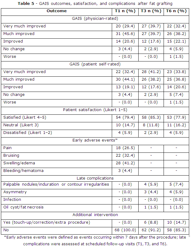
GAIS-based aesthetic assessment showed that the proportions rated as much improved and very much improved predominated at all follow-up time points. According to physician assessment, the proportion rated much/very much improved was 75.0% at T1, 79.4% at T3, and 70.6% at T6. Based on patient self-assessment, the corresponding rates were 76.5% at T1, 79.4% at T3, and 70.6% at T6. Patient satisfaction (Likert 4–5) was 79.4% at T1, increased to 85.3% at T3, and remained 77.9% at T6 (table 5).
Regarding safety, early adverse events (≤ 7 days) mainly included swelling/edema (41.2%), bruising (32.4%), pain (26.5%), and bleeding/hematoma (4.4%), with no events recorded at T3 and T6. Late complications (> 7 days) were primarily palpable nodules/induration or surface contour irregularities (5.9% at T3; 7.4% at T6) and asymmetry (4.4% at T3; 5.9% at T6). No infection was observed; oil cyst/fat necrosis was 1.5% at T3 and persisted at 1.5% at T6. The need for additional interventions increased over time: 0% at T1, 8.8% at T3, and 14.7% at T6 (table 5).
DISCUSSION
Characteristics of the study population and baseline nasolabial fold severity
In this study, the mean age of participants was 39.1 ± 6.9 years, and most were women (89.7%). This age group commonly seeks minimally invasive facial rejuvenation and still has favorable potential for improvement in skin quality and soft-tissue structure. Baseline nasolabial fold depression was predominantly moderate to severe according to the WSRS (mean scores: Left 3.32 ± 0.47; right 3.34 ± 0.47), consistent with indications for volumization to improve the nasolabial folds. Notably, the proportion of participants with a history of facial aesthetic procedures was high (80.9%). This factor may influence tissue characteristics (e.g., fibrosis/scarring, altered fat compartments, and dermal–subdermal changes), which in turn can affect graft take and the stability of outcomes after fat grafting. Therefore, standardizing injection technique by plane, using micro-aliquots, and conducting longitudinal follow-up are important for accurately evaluating real-world effectiveness.
Improvements based on WSRS and GAIS
Current findings demonstrated a marked reduction in WSRS on both sides after fat grafting, with the strongest improvement during the 1–3 month period and maintenance through 6 months, although with a slight increase compared with the 3-month assessment. This pattern is consistent with the biology of fat grafting: The early phase is influenced by postoperative edema, revascularization, and stabilization of the graft, whereas partial volume reduction may occur later due to remodeling and replacement of transplanted adipose tissue by fibrosis to varying degrees across individuals, leading to a mild decline in effect over time. Carruthers KH et al.(8) reported substantial variability in volume retention after low-volume facial fat grafting, emphasizing the influence of processing technique on outcomes. Overall, the current evidence indicates that fat-graft retention remains variable across different techniques and follow-up intervals.
GAIS assessment similarly indicated favorable aesthetic improvement: The proportion rated as much improved/very much improved increased from 75.0% at 1 month to 79.4% at 3 months and remained 70.6% at 6 months (physician rated). The peak effect at 3 months may reflect a time point at which graft stabilization is optimal and postoperative swelling has largely resolved, resulting in a more natural contour of the nasolabial fold. Combining WSRS (a relatively objective fold-severity measure) with GAIS (a global aesthetic improvement assessment) provides a more multidimensional description of treatment effectiveness in aesthetic practice, particularly when the goal is not only to reduce fold depth but also to improve midface harmony.
When comparing autologous fat grafting with dermal filler techniques, differences in mechanisms and durability should be considered. A systematic review and meta-analysis by Stefura T et al.(4) on fillers for nasolabial folds (including randomized clinical trials) reported pooled WSRS reductions from 3.23 before treatment to 1.79 at 1 month, 2.02 at 6 months, and 2.46 at 12 months, suggesting waning effects over time and a tendency toward repeat treatments to maintain optimal outcomes. In a randomized, double-blind, split-face trial involving 74 individuals with moderate-to-severe nasolabial folds, Kim BR et al.(5) reported a reduction in WSRS from 3.16 ± 0.84 to 2.59 ± 0.87 at week 24 after hyaluronic acid filler injection, with mainly mild and transient local reactions. Thus, autologous fat grafting offers advantages in volume restoration and the potential to improve skin quality, but it is also characterized by variability in fat retention and a non-negligible proportion of patients requiring secondary interventions.
Patient satisfaction and need for additional interventions
Patient satisfaction peaked at 3 months (satisfied/very satisfied: 85.3%) and declined slightly at 6 months (77.9%). This trend parallels the temporal changes observed in WSRS/GAIS, suggesting that minor volume regression over time may directly influence patients’ aesthetic perception. The proportion requiring additional intervention/touch-up increased gradually to 14.7% at 6 months, reflecting clinical practice in which facial fat grafting often benefits from a staged approach, particularly in patients with deeper folds or suboptimal baseline tissue quality, to optimize smoothness and durability of outcomes. Evidence on the durability of injectables also indicates the need for maintenance over time; for dermal fillers, a systematic review reported a notable number of adverse events and unwanted reactions across follow-up studies, although most were mild to moderate highlighting that expectation management and follow-up/retreatment planning are integral to aesthetic treatment strategies.(6)
Safety and complications
With respect to safety, early adverse events in our study were mainly pain (26.5%), bruising (32.4%), and prolonged swelling/edema (41.2%). These are common local reactions after harvest-and-inject procedures and generally resolve with appropriate care. Importantly, late complications were infrequent: Palpable nodules/induration or contour irregularities (7.4% at 6 months), asymmetry (5.9% at 6 months), and oil cyst/fat necrosis at 1.5%.
Compared with the literature, Schiraldi L et al.(9) (systematic review of complications of facial fat grafting) reported wide variability in complication rates across studies and categorized adverse effects from mild to severe, including rare but serious events related to intravascular injection or fat embolization, which require specialized management and may result in long-term sequelae. In addition, a pooled analysis by Moellhoff N et al.(15) of cases with arterial occlusion after facial fat grafting (61 patients) reported a high rate of permanent visual impairment and, in some cases, concomitant central nervous system injury, emphasizing that although uncommon, vascular complications represent a severe risk that must be prevented through strict adherence to safe injection techniques and appropriate selection of injection planes.
In current study, no severe vascular events were recorded. This may be related to the use of blunt cannulas, tunnel creation, micro-aliquot placement with layered distribution, and early postoperative monitoring. Nevertheless, because severe events are rare and highly dependent on technical execution and high-risk vascular anatomy, continued standardization of safety protocols and anatomy-based training for facial injection techniques remain essential, particularly in zones with higher risk of intravascular injection.
This study is strengthened by systematic recording of processing parameters and side-specific injection volumes, enabling future analyses of predictors of response and adverse events. Other strengths include longitudinal follow-up at 1, 3, and 6 months, use of widely adopted scales (WSRS, GAIS), and concurrent reporting of effectiveness, satisfaction, complications, and touch-up needs. Limitations include the lack of a control group and a 6-month follow-up that may not reflect long-term durability. Outcomes were mainly based on subjective clinical scales without objective volumetric assessment (ultrasound). Future studies should extend follow-up to ≥ 12 months, incorporate objective volumetry, and develop predictive models considering prior procedures, tissue factors, weight change, layer-specific volumes.
In conclusion, autologous fat grafting for the treatment of nasolabial fold depression in this study resulted in meaningful improvement on WSRS and GAIS, peaking at 3 months and maintained through 6 months with a slight decline over time; patient satisfaction was high and late complications were uncommon. In clinical practice, these findings support autologous fat grafting as an effective and safe option when strict technical and injection-safety principles are followed. Pre-procedural counseling should address the variability in fat retention and the potential need for touch-up injections to optimize long-term aesthetic stability.
BIBLIOGRAPHIC REFERENCES
1. Quan Y, Zhang T, Liu L, Han B, Cao Z, Shen Y. Quantitative Assessment of Nasolabial Fold Characteristics Across Age Groups [Internet]. Aesthetic Surgery Journal Open Forum. 2025; 7:1-13. DOI: 10.1093/asjof/ojaf075
2. Hung WK, Chen CB, Cheng CY, Chang SL, Hu S, Chang YC, et al. The aging process of deep fat compartments in the midface and midfacial rejuvenation: An ultrasound-based analysis [Internet]. Dermatologica Sinica. 2023; 41(4):251-256. DOI: 10.4103/ds.DS-D-23-00179
3. Day DJ, Littler CM, Swift RW, Gottlieb S. The Wrinkle Severity Rating Scale [Internet]. American Journal of Clinical Dermatology. 2004; 5(1):49-52. DOI:10.2165/00128071-200405010-00007
4. Stefura T, Kacprzyk A, Droś J, Krzysztofik M, Skomarovska O, Fijałkowska M, et al. Tissue Fillers for the Nasolabial Fold Area: A Systematic Review and Meta-Analysis of Randomized Clinical Trials [Internet]. Aesthetic Plastic Surgery. 2021; 45(5):2300-16. DOI: 10.1007/s00266-021-02439-5
5. Kim BR, Shin J-W, Kim D-W, Choung JJ, Jang Y, Lee S-H, et al. Efficacy and Safety of New Hyaluronic Acid Filler for Nasolabial Fold Correction: A Double-Blind, Randomized Trial [Internet]. Plastic and Reconstructive Surgery. 2026; 157(2):249-56. DOI: 10.1097/prs.0000000000012320
6. Janovskiene A, Chomicius D, Afanasjevas D, Petronis Z, Razukevicius D, Jagelaviciene E. Safety and Potential Complications of Facial Wrinkle Correction with Dermal Fillers: A Systematic Literature Review [Internet]. Medicina. 2025; 61(1):25. DOI: 10.3390/medicina61010025
7. Vasavada A, Raggio BS. Autologous fat grafting for facial rejuvenation. In StatPearls [Internet]. StatPearls Publishing; 2023. [access: 21/01/2026]. Available from: https://www.ncbi.nlm.nih.gov/books/NBK557860/
8. Carruthers KH, Austen WG, Jr, Remy K, Hamaguchi R, Liu S, Vyas K, et al. Improving the Retention of Low-Volume Autologous Fat Grafting: A Comparative Analysis of Lipoaspirate Processing Techniques for Facial Feminization [Internet]. Aesthetic Surgery Journal Open Forum. 2024; 6:1-8. DOI: 10.1093/asjof/ojae043
9. Schiraldi L, Sapino G, Meuli J, Maruccia M, Cherubino M, Raffoul W, et al. Facial Fat Grafting (FFG): Worth the Risk? A Systematic Review of Complications and Critical Appraisal [Internet]. Journal of Clinical Medicine. 2022; 11(16):4708. DOI: 10.3390/jcm11164708
10. Brown K, McCoskey M, Nakra T. Complications of facial autologous fat grafting [Internet]. Plastic and Aesthetic Research. 2024; 11(7):8-14. DOI: 10.20517/2347-9264.2023.94
11. Chaghamirzayi P, Abdi H, Rozveh JK, Nejad MA, Azizmanesh M. Fat embolism following fat grafting: A systematic review of reported cases. [Internet]. JPRAS Open. 2025; 43:18-55. DOI: 10.1016/j.jpra.2024.10.012
12. Egro FM, Roy E, Rubin JP, Coleman SR. Evolution of the Coleman technique [Internet]. Plastic and Reconstructive Surgery. 2022; 150(2):329-36. DOI: 10.1097/PRS.0000000000009355
13. Shauly O, Gould DJ, Ghavami A. Fat grafting: basic science, techniques, and patient management [Internet]. Plastic and Reconstructive Surgery–Global Open. 2022; 10(3):e3987. DOI: 10.1097/GOX.0000000000003987
14. Venkataram J. Tumescent liposuction: a review [Internet]. Journal of cutaneous and aesthetic surgery. 2008; 1(2):49-57. DOI: 10.4103/0974-2077.44159
15. Moellhoff N, Kuhlmann C, Frank K, Kim BS, Conte F, Cotofana S, et al. Arterial Embolism After Facial Fat Grafting: A Systematic Literature Review [Internet]. Aesthetic Plastic Surgery. 2023; 47(6):2771-87. DOI: 10.1007/s00266-023-03511-y
Conflicts of interest
The authors declare no conflict of interest.
Funding
No external funding was received for this study.
Authorship contribution
Conceptualization: Nguyen Van Phung, Nguyen Thanh Van, Le Minh Phong.
Data curation: Nguyen Van Phung, Le Minh Phong, Nguyen Thanh Van.
Formal analysis: Nguyen Van Phung, Le Minh Phong.
Research: Nguyen Van Phung, Le Minh Phong.
Methodology: Nguyen Van Phung, Nguyen Thanh Van, Phan Hung Phuc.
Project administration: Nguyen Van Phung, Nguyen Thanh Van.
Supervision: Nguyen Thanh Van, Le Minh Phong, Phan Hung Phuc.
Validation: Nguyen Van Phung, Nguyen Thanh Van.
Drafting - Revision and editing: Nguyen Van Phung, Nguyen Thanh Van, Phan Hung Phuc.
Data Availability
This research data is confidential according to the applicable confidentiality agreements and regulations and, therefore, cannot be publicly displayed or shared. Access to these data requires proper authorization. For any questions or further information, please contact drthanhvan@gmail.com